Throughout your pregnancy, you’ll have multiple ultrasounds. Most people have at least one early on, which is used (among other things) for pregnancy dating. It’s also common to have an ultrasound at the end of pregnancy to estimate fetal size. By far the longest ultrasound will come around the 20th week of pregnancy, in what is called an anatomy scan.
Generally: this scan is a way to check the full fetal anatomy at this stage of pregnancy and to either confirm that everything looks good or possibly raise concerns that need follow-up. Most of the time, this experience is straightforward, simple, and reassuring. But it’s also medically important. If something is wrong, you want to know.
The anatomy scan differs from other ultrasounds only in scope. It will be a lot longer than your other ultrasounds, and they’ll take a lot more measurements. And yes, there will be pictures.
What to expect at the anatomy scan
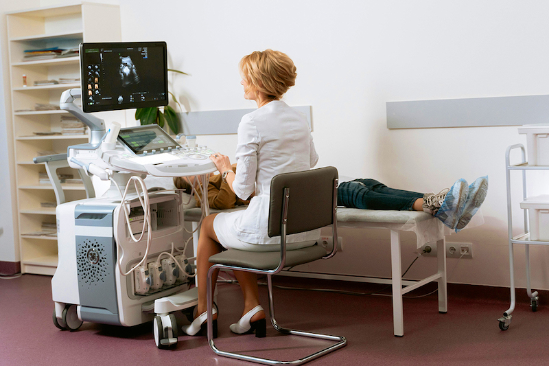
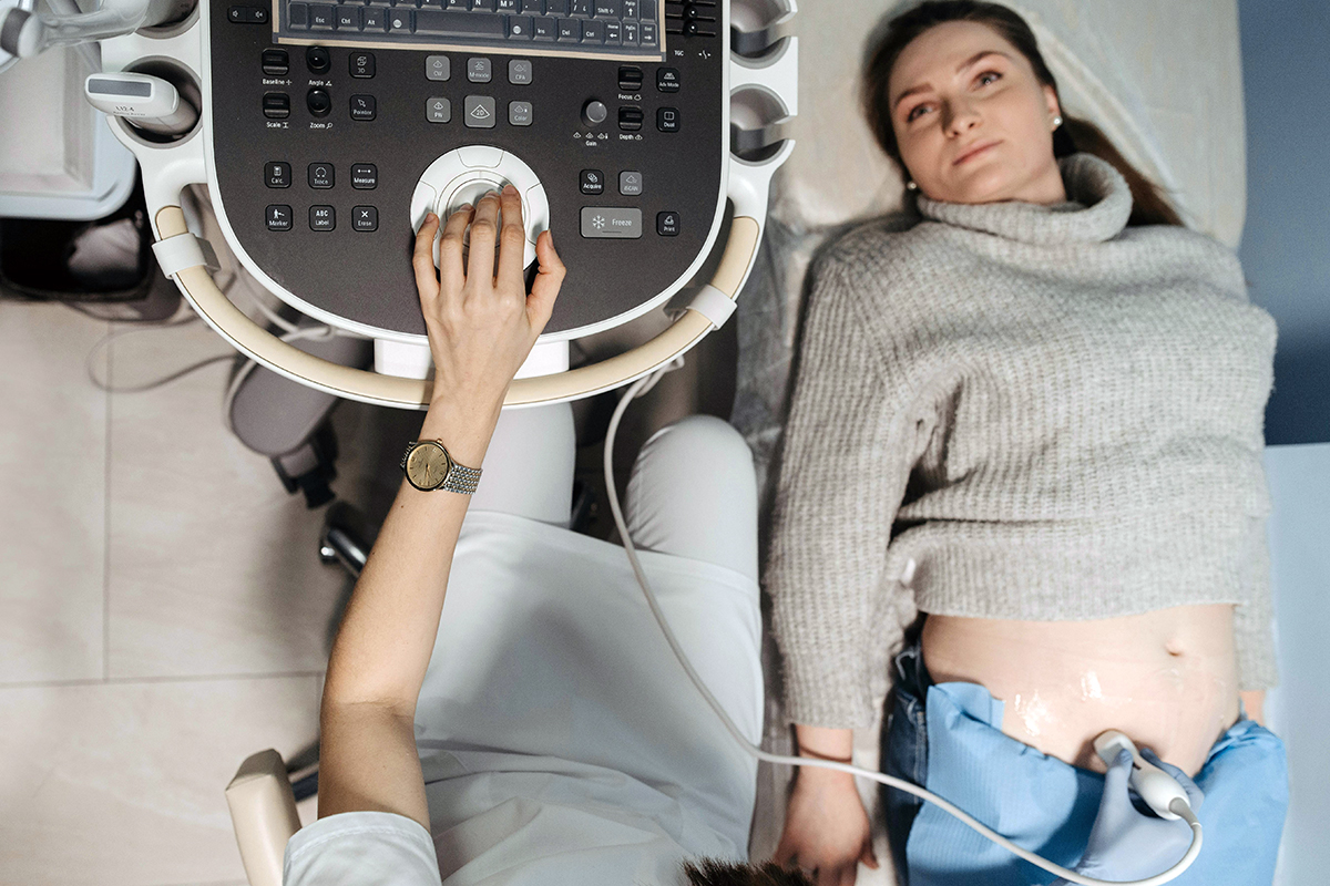
At this 20-week scan (which typically occurs, in fact, sometime between 18 and 22 weeks), you’ll have an ultrasound for about 45 minutes. You don’t need to prepare for it, although your provider may recommend you have a full bladder since this can make imaging easier.
The ultrasound tech will be focused on scanning and measuring many features of the fetus. They’ll take pictures and click around. It’s often very quiet — with one hand they move the wand, then they take a picture (keyboard click) and then measure an organ (click, click) and then move the wand again and repeat. This can be oddly unnerving — wait! Is that a bad click? Does that number look right? — but it’s all typical. Your ultrasound technician will not comment, so don’t expect much of an interactive experience. Their job is to take the measurements and send them to your provider to interpret.

This anatomy scan focuses on virtually all body parts. The ultrasound tech will measure things like the liver and kidneys, will look at how the heart is structured and how blood is flowing through it, at fingers and toes, legs, head, sex organs, and so on. They will also take some more general measurements that tell them whether the fetus is, size-wise, where you’d expect it to be.
The ultrasound also lets doctors see amniotic fluid levels and the placement of the placenta.
After the anatomy scan, you will generally have an appointment with your doctor (either at the same visit or soon after) to discuss the results. They can go over the findings with you and discuss concerns, if any came up.
What can I learn from it?
Issues with the fetus
The anatomy scan is looking for possible anatomical abnormalities with the fetus. These issues could be markers of another condition or they could be issues on their own. An example in the first category: there are some signs in the scan of the heart that are “soft markers” for Down syndrome. If a fetus has these markers, it is more likely they have Down syndrome. An example in the second category is spina bifida, a condition in which the spinal cord does not form properly. This can often be visualized in the anatomy scan.
Other issues that could be detected include missing kidneys, issues with the intestine or abdominal walls, cleft lip, or skeletal issues. These are scary, but it’s important to say that they are rare.
The information from the anatomy scan also needs to be taken in context with what you know before. Our technology for detecting chromosomal abnormalities has improved tremendously over time, and many people will have had cell-free fetal DNA testing earlier in their pregnancy that will have ruled out some of these issues. In terms of testing for conditions like Down syndrome and trisomy 13 and 18, the earlier testing is much more accurate than what you’d learn from the markers detected in this scan. This means that even if the ultrasound tech saw something that could suggest an increased risk of a chromosomal issue, you would have ruled that out.
If there is an issue detected with the fetus, your doctor will talk you through next steps. Usually, more follow-up testing or imaging would be needed to learn more and consider what might be the right course of action.
Other pregnancy concerns
There are two other issues that might be detected during this scan. One is placenta previa, where the placenta either fully or partially covers the cervix. If this persists until full term, you will need a caesarean section. However, about 90% of cases of previa diagnosed at 20 weeks resolve before full term (as the uterus grows, the placenta moves). This is not something to panic about. If it is diagnosed, your doctor may suggest restrictions on sexual activity.
The other issue is either too little or too much amniotic fluid. Either of these conditions — called oligohydramnios and polyhydramnios — can occur on their own or as a sign of a broader issue. If you have either one, your doctor will work to rule out possible fetal causes. If these are ruled out, they will continue to monitor the fluid levels.
Fetal sex
You can learn fetal sex at this ultrasound, if you have not already learned it through other testing. At this stage of pregnancy, fetal sex is nearly always clearly visible. This means if you do not want to know, you should be very clear with your ultrasound tech and doctor up front so they do not accidentally reveal it.
Other common questions
Do I have to have this scan?
This is a routine scan for nearly everyone, but not every doctor will insist on it, especially if you have had other testing that would detect many conditions. In my second pregnancy, since I had both a cell-free fetal DNA test and an amniocentesis, my midwife and I agreed there wasn’t much we would learn from this ultrasound, so I skipped it. Having said that, since this test isn’t invasive and doesn’t carry any risks (and produces good baby pictures!), most people are happy to have it.
Are ultrasounds risky?
Over the years, concerns have been raised at various points about the use of ultrasound. Some speculation has linked them to higher rates of autism or other developmental disorders. This is just that: speculation. There is no evidence or data to support a risk.
The only connection with any evidence — and this is quite weak — is a possible link between ultrasounds and left-handedness. The effect is very small, not statistically precise, and this outcome isn’t an important one.
Overall: there is no reason to avoid ultrasounds, and there are many reasons to have them.
Is there a difference between 2D, 3D, and 4D ultrasounds?
These are all ultrasounds, which use different imaging approaches to visualize in more or less detail. Standard ultrasounds are flat — 2D — like a photo. A 3D ultrasound allows you to see contours of the baby. A 4D ultrasound is, basically, a video. For the anatomy scan, a 2D ultrasound is standard. If there is a concern with the fetus, your doctor may use a 3D or 4D scan to learn more. Generally, though, these are not necessary.
The bottom line
- The anatomy scan, usually done between 18 and 22 weeks of pregnancy, is a detailed ultrasound that focuses on the fetus’s full anatomy.
- During the ultrasound, a technician will check the fetus for potential abnormalities, measure organ structures, and assess growth milestones. They also evaluate the pregnant woman’s amniotic fluid levels and placenta placement.
- Cell-free fetal DNA testing, often done earlier in pregnancy, can complement and help contextualize findings from the anatomy scan.
- Fetal sex can often be clearly identified during this scan.


Log in
I just want to mention that the ultrasound may not be very long. I’m in my second pregnancy and both times (at two different practices) the ultrasound was less than 30min. I remember being very freaked out the first time because I’d only heard about people having really long (think two hour) anatomy scans.